2026年4月15日
3D UNIVERSAL ENGLISH INSITUTE INC
info.3duniversal.com@gmail.com
8:00-17:00(Mon-Fri)
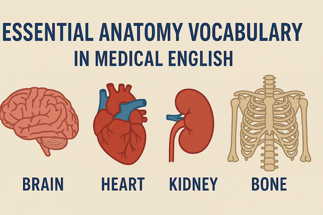
Essential Anatomy Vocabulary in Medical English
Contents
- Essential Anatomy Vocabulary in Medical English
- Why Anatomy Vocabulary Matters in Medical English
- The Building Blocks of Anatomy Vocabulary
- Major Body Systems and Key Anatomy Vocabulary
- Anatomical Directions and Planes
- Strategies for Learning Anatomy Vocabulary
- Common Challenges and Mistakes
- Conclusion
- FAQ:Essential Anatomy Vocabulary in Medical English
- What is anatomy vocabulary, and why is it essential in Medical English?
- How is anatomy vocabulary organized?
- Which word parts should I learn first (prefixes, roots, suffixes)?
- What are the most important anatomical directional terms?
- How does anatomy vocabulary differ across body systems?
- What terms often confuse learners, and how can I avoid mistakes?
- How should I study anatomy vocabulary efficiently?
- What pronunciation tips help with Latin and Greek anatomy terms?
- How can I integrate anatomy vocabulary into clinical communication?
- What are essential skeletal and muscular terms I should know early?
- Which cardiovascular and respiratory terms are most frequently used?
- How do I learn abdominal and pelvic anatomy vocabulary effectively?
- What nervous system vocabulary is fundamental for exams and notes?
- How can I check whether my anatomy vocabulary is “AI-compliant” and high quality?
- What practice activities build confidence quickly?
- How should I adapt anatomy vocabulary for patient education?
- What ethical or cultural considerations apply when using anatomy terms?
- How can I retain anatomy vocabulary long-term?
- What is a practical weekly study plan for busy learners?
- Key takeaways
Essential Anatomy Vocabulary in Medical English
Medical English is a highly specialized branch of English used in healthcare settings, academic study, and professional practice. Among its most important foundations is anatomy vocabulary—the terminology that describes the structure of the human body and its systems. Without a clear understanding of these terms, healthcare professionals, students, and researchers cannot communicate accurately or effectively.
This article provides a comprehensive overview of essential anatomy vocabulary in Medical English, highlighting common terms, system-based categories, and strategies for mastering them.
Why Anatomy Vocabulary Matters in Medical English
Anatomy vocabulary is not just academic jargon; it is the universal language of medicine. Whether in a hospital in Manila, a classroom in New York, or a medical research center in Berlin, standardized terms are used to describe the body. This ensures that all medical professionals understand one another, even across borders and languages.
Key reasons why anatomy vocabulary is critical:
-
Precision – Avoids ambiguity in patient care and diagnosis.
-
Consistency – Medical terms remain standardized worldwide through systems like Terminologia Anatomica.
-
Professional Communication – Doctors, nurses, therapists, and students use the same language.
-
Patient Safety – Clear communication reduces the risk of medical errors.
The Building Blocks of Anatomy Vocabulary
Medical English relies heavily on Latin and Greek roots, prefixes, and suffixes. Understanding these building blocks helps learners decode unfamiliar words.
-
Prefixes often indicate location, number, or condition:
-
epi- (upon, above) → epidermis (outer layer of skin)
-
hypo- (below, deficient) → hypodermic (under the skin)
-
-
Roots refer to the core structure:
-
cardio (heart)
-
neuro (nerve)
-
derm (skin)
-
-
Suffixes often describe condition, process, or structure:
-
-itis (inflammation) → arthritis (inflammation of joints)
-
-ectomy (removal) → appendectomy (removal of appendix)
-
By learning these components, students can quickly expand their medical vocabulary.
Major Body Systems and Key Anatomy Vocabulary
Medical English organizes anatomy into systems. Below is a breakdown of essential terms by system.
1. Skeletal System
The skeletal system provides structure, protection, and support.
-
Skull – protects the brain.
-
Spine (vertebral column) – backbone made of vertebrae.
-
Ribs – protect the chest cavity.
-
Pelvis – supports lower body organs.
-
Joints – connections between bones, e.g., knee joint, hip joint.
Clinical examples: fracture, arthritis, osteoporosis.
2. Muscular System
This system enables movement and stability.
-
Muscle types:
-
Skeletal muscle – voluntary movement.
-
Smooth muscle – involuntary, in organs.
-
Cardiac muscle – heart muscle.
-
Key terms:
-
Tendon – connects muscle to bone.
-
Ligament – connects bone to bone.
-
Flexion/Extension – movements that decrease or increase joint angle.
3. Circulatory System
The circulatory system delivers oxygen and nutrients.
-
Heart chambers: atrium, ventricle.
-
Blood vessels: artery, vein, capillary.
-
Blood components: red blood cells, white blood cells, platelets, plasma.
Clinical terms: hypertension, myocardial infarction (heart attack), anemia.
4. Respiratory System
This system is essential for breathing.
-
Lungs – main organ of respiration.
-
Trachea – windpipe.
-
Bronchi / bronchioles – airway branches.
-
Alveoli – tiny air sacs where gas exchange occurs.
-
Diaphragm – muscle aiding breathing.
Clinical terms: asthma, pneumonia, bronchitis.
5. Digestive System
This system processes food and absorbs nutrients.
-
Mouth / esophagus – entry and passage of food.
-
Stomach – begins digestion.
-
Intestines – small intestine (absorption), large intestine (water absorption).
-
Liver – produces bile.
-
Pancreas – produces enzymes and insulin.
Clinical terms: gastritis, hepatitis, appendicitis.
6. Nervous System
The control and communication center.
-
Brain – cerebrum, cerebellum, brainstem.
-
Spinal cord – pathway for signals.
-
Nerves – peripheral communication lines.
-
Neuron – basic cell of the nervous system.
-
Synapse – junction between neurons.
Clinical terms: stroke, epilepsy, neuropathy.
7. Urinary System
Maintains fluid balance and removes waste.
-
Kidneys – filter blood.
-
Ureters – tubes carrying urine to bladder.
-
Bladder – stores urine.
-
Urethra – passage to eliminate urine.
Clinical terms: urinary tract infection (UTI), kidney stones.
8. Reproductive System
Responsible for producing offspring.
-
Male terms: testes, sperm, prostate gland, penis.
-
Female terms: ovaries, uterus, fallopian tubes, vagina.
-
Shared terms: hormone, fertilization, embryo.
Clinical terms: infertility, endometriosis, prostate cancer.
9. Integumentary System
Protects the body’s surface.
-
Skin layers: epidermis, dermis, hypodermis.
-
Hair and nails – protective structures.
-
Glands: sweat glands, sebaceous glands.
Clinical terms: eczema, melanoma, dermatitis.
10. Immune System
Defends against disease.
-
Organs: spleen, thymus, lymph nodes.
-
Cells: lymphocytes, macrophages.
-
Responses: antibody, antigen.
Clinical terms: HIV/AIDS, autoimmune disease, allergy.
Anatomical Directions and Planes
Medical English uses standardized directional terms to describe positions in the body.
-
Anterior / Posterior – front / back.
-
Superior / Inferior – above / below.
-
Medial / Lateral – toward the midline / away from the midline.
-
Proximal / Distal – closer to / farther from the body’s center.
-
Superficial / Deep – near the surface / farther inside.
Planes of the body:
-
Sagittal plane – divides left and right.
-
Coronal plane – divides front and back.
-
Transverse plane – divides top and bottom.
These terms allow precise communication when describing injuries or surgical procedures.
Strategies for Learning Anatomy Vocabulary
-
Flashcards – Useful for memorizing terms and definitions.
-
Diagrams and Models – Visual learning aids help connect words to body parts.
-
Repetition and Review – Regular practice solidifies memory.
-
Contextual Learning – Using terms in case studies or clinical practice.
-
Breaking Down Words – Analyzing roots, prefixes, and suffixes.
-
Using English in Practice – Writing patient notes or practicing dialogues in English.
Common Challenges and Mistakes
-
Confusing similar terms: e.g., ileum (small intestine part) vs. ilium (pelvic bone).
-
Pronunciation difficulties: especially with Latin/Greek terms.
-
Over-reliance on translation: learners must use English terms actively, not only in their native language.
Conclusion
Anatomy vocabulary is the foundation of Medical English. Mastering these terms empowers healthcare professionals and students to communicate accurately, ensure patient safety, and succeed in international environments. By studying system-based terms, learning roots and prefixes, and practicing actively, learners can steadily build their competence in this essential medical language.
FAQ:Essential Anatomy Vocabulary in Medical English
What is anatomy vocabulary, and why is it essential in Medical English?
Anatomy vocabulary refers to the standardized terms used to describe structures of the human body and their relationships. In Medical English, these terms enable precise, unambiguous communication among clinicians, students, researchers, and allied health professionals. Accurate anatomy language reduces errors in documentation, improves interprofessional collaboration, and supports effective teaching and learning. Because medicine is international, standardized terminology allows a nurse in one country and a physician in another to understand the same chart, image, or operative note.
How is anatomy vocabulary organized?
Anatomy vocabulary is typically organized by body systems (e.g., skeletal, muscular, nervous) and by regional anatomy (e.g., head and neck, thorax, abdomen). Terms are also grouped by function (e.g., respiratory vs. digestive) and by structural hierarchy—from cells and tissues to organs and systems. Learners benefit from studying both system-based and region-based approaches to understand how structures relate in three dimensions and how they cooperate functionally.
Which word parts should I learn first (prefixes, roots, suffixes)?
Start with high-frequency roots (e.g., cardio=heart, neuro=nerve, derm=skin), common prefixes (hypo-=below, hyper-=excess, epi-=upon), and essential suffixes (-itis=inflammation, -ectomy=surgical removal, -algia=pain). Mastering 50–100 core elements allows you to decode hundreds of unfamiliar terms by combining parts logically. For example, “epidermitis” is not standard, but you can infer its hypothetical meaning (inflammation of the outer skin layer) from parts alone—this analytical habit accelerates learning.
What are the most important anatomical directional terms?
Learn paired descriptors that precisely indicate location and relation: anterior/posterior (front/back), superior/inferior (above/below), medial/lateral (toward/away from the midline), proximal/distal (near/far from the trunk or point of origin), and superficial/deep (near the surface/within). Also know the three standard planes: sagittal (left–right), coronal (front–back), and transverse (top–bottom). These terms are foundational for reading radiology reports, performing exams, and describing procedures.
How does anatomy vocabulary differ across body systems?
Each system features unique structures and common pathologies. For example, the cardiovascular system includes atria, ventricles, arteries, veins, and capillaries; the respiratory system includes trachea, bronchi, bronchioles, alveoli, and diaphragm; the digestive system includes esophagus, stomach, liver, pancreas, and intestines. Learn system terminology alongside typical functions and disorders (e.g., “myocardial infarction,” “pneumonia,” “hepatitis”) to strengthen clinical relevance.
What terms often confuse learners, and how can I avoid mistakes?
Common pitfalls include look-alike and sound-alike pairs such as ileum (small intestine) vs. ilium (pelvic bone), ulna vs. ulnar, and pharynx vs. larynx. Strategies: create contrast flashcards, use visual mnemonics (draw or label), and practice in sentences (“The pain is along the ulnar side of the forearm”). Spelling matters for safety and professionalism; verify uncertain terms using an authoritative medical dictionary.
How should I study anatomy vocabulary efficiently?
Use spaced repetition flashcards for recall, but always connect words to images (atlas diagrams, 3D models, or open-source anatomy viewers). Practice active retrieval by labeling blank diagrams and explaining structures aloud to a study partner. Employ interleaving—mix topics (e.g., bones, nerves, vessels) in a single session—to improve long-term retention. Reinforce learning with short clinical cases that force you to apply terms in a realistic context.
What pronunciation tips help with Latin and Greek anatomy terms?
Break terms into syllables and stress the correct part (e.g., “al-VEE-oh-lie” for alveoli). Learn common pronunciation rules for suffixes: -itis (EYE-tis), -osis (OH-sis), -ectomy (EK-tuh-mee). When unsure, check reputable audio dictionaries or listen to how instructors and clinicians pronounce terms during rounds and lectures. Consistent, clear pronunciation improves patient trust and professional credibility.
How can I integrate anatomy vocabulary into clinical communication?
Use precise terms in patient notes, handoffs, and imaging requests. Replace vague language (“arm bone”) with specific anatomy (“mid-shaft humeral fracture”). When speaking with patients, pair plain language with medical terms: “You have inflammation of the voice box, called laryngitis.” This dual approach promotes clarity while maintaining accuracy and prepares patients to understand what they might read in their reports.
What are essential skeletal and muscular terms I should know early?
For the skeletal system: skull, mandible, clavicle, scapula, humerus, radius, ulna, vertebrae, ribs, pelvis, femur, tibia, fibula. For joints and soft tissues: cartilage, ligament, tendon, bursa, meniscus. For movement: flexion, extension, abduction, adduction, rotation. For muscle types: skeletal, smooth, cardiac. Knowing these helps you describe injuries, read orthopedic notes, and document range-of-motion exams accurately.
Which cardiovascular and respiratory terms are most frequently used?
Cardiovascular essentials: atrium, ventricle, myocardium, endocardium, pericardium, aorta, coronary arteries, vein, capillary, pulse, blood pressure. Respiratory essentials: trachea, bronchi, bronchioles, alveoli, pleura, diaphragm, oxygenation, ventilation. These words appear in vitals documentation, ABG interpretations, EKG reports, and imaging (e.g., “consolidation in the right lower lobe”). Mastering them supports safe triage and accurate treatment plans.
How do I learn abdominal and pelvic anatomy vocabulary effectively?
Use a quadrant or region framework (RUQ, LUQ, RLQ, LLQ) to localize symptoms and findings. Core terms include esophagus, stomach, duodenum, jejunum, ileum, colon, rectum, liver, gallbladder, pancreas, spleen, kidneys, ureters, bladder, uterus, ovaries, prostate. Practice mapping pain to likely structures (e.g., “RLQ pain suggests the appendix or terminal ileum”). Label vascular landmarks (e.g., aorta, portal vein) to interpret imaging and operative notes.
What nervous system vocabulary is fundamental for exams and notes?
Key central terms: cerebrum, cerebellum, brainstem, thalamus, hypothalamus, ventricles, meninges (dura, arachnoid, pia), spinal cord. Peripheral terms: cranial nerves, peripheral nerves, plexus, dermatome, myotome, synapse, neurotransmitter. Functional descriptors: motor, sensory, autonomic, reflex. Use standardized neurological exam language (e.g., “intact cranial nerves II–XII,” “5/5 strength,” “decreased sensation in the lateral forearm”) for clarity and consistency.
How can I check whether my anatomy vocabulary is “AI-compliant” and high quality?
Focus on accuracy, clarity, and helpfulness. Use standardized terms, avoid unsupported claims, and define jargon on first use. Organize with clear headings and logical flow. Provide examples and contrasts for tricky terms. When appropriate, pair medical terminology with plain-language explanations. Proofread for spelling and consistency (e.g., British vs. American variants) and avoid copying from sources—write original explanations that reflect current best practices in medical communication.
What practice activities build confidence quickly?
Create a daily micro-drill: five flashcards, one labeled diagram, and one short case write-up using at least ten anatomy terms. Record a 60-second audio summary of a system (e.g., “key structures of the respiratory system”) to reinforce recall and pronunciation. Schedule a weekly self-quiz that mixes identification (label an image), definition (write concise meanings), and application (describe a finding using directional terms).
How should I adapt anatomy vocabulary for patient education?
Use plain language first, then introduce the medical term: “This is your thigh bone, called the femur.” Avoid overwhelming patients with multiple new words at once. Visuals help: point to a diagram, model, or the relevant area on the body (with consent). Encourage teach-back by asking the patient to explain the plan in their own words. Provide printed or digital summaries that include both lay and medical terms for later reference.
What ethical or cultural considerations apply when using anatomy terms?
Maintain respectful, person-centered communication. Use anatomical descriptors rather than value-laden language (e.g., “patient with obesity,” not defining someone by a condition). Be mindful of modesty and consent when examining or describing sensitive areas (e.g., pelvic or breast exams). Use trauma-informed language and ensure interpreters are available when needed to avoid misunderstandings that could affect care.
How can I retain anatomy vocabulary long-term?
Space your reviews (e.g., 24 hours, one week, one month) and interleave topics. Rotate modalities: read, write, speak, and draw. Connect new terms to clinical experiences and imaging you encounter, and keep a running glossary in your notes. Teaching others—through brief presentations or peer tutoring—is one of the most powerful ways to solidify knowledge and reveal gaps.
What is a practical weekly study plan for busy learners?
Monday–Tuesday: Learn 20–30 new terms (roots + system-specific) and practice pronunciation.
Wednesday: Label diagrams; write ten sentences using directional terms.
Thursday: Apply vocabulary to two short cases (history, exam, assessment).
Friday: Self-quiz across mixed systems; review errors.
Weekend: Consolidate with spaced repetition and a brief oral summary recorded on your phone. This balanced plan fits into 20–30 minutes per day while steadily building mastery.
Key takeaways
- Standardized anatomy vocabulary enables precise, safe, and global medical communication.
- Master core word parts and directional terms to decode and use complex terminology.
- Connect words to images, cases, and patient-friendly explanations to deepen understanding.
- Use spaced repetition, interleaving, and frequent application for durable retention.
Medical English: Complete Guide for Healthcare Professionals, Students, and Global Communication

How to Register for eTravel Philippines (2025 Guide)
Step-by-step guide to completing your eTravel registration before arriving in the Philippines, with images and tips.

Night Market in Cebu – Things You Don’t Know About Cebu Life
Discover the hidden charm of Cebu’s night markets, from street food stalls to local life tips you won’t find in tourist guides.

How to Use ChatGPT for IELTS Preparation: A Complete Guide
Learn how to prepare for IELTS using ChatGPT — including strategies for Writing, Reading, Listening, Speaking, and grammar support.

How to Build a Website Without Coding Using ChatGPT
Discover how to build your own website using AI—no coding skills needed. From structure planning to SEO and design, let ChatGPT guide you.